why anticoagulants are contraindicated in portal vein thrombosis ?


Anticoagulants are drugs that help prevent the formation of blood clots or dissolve existing blood clots. However, in the case of portal vein thrombosis (PVT), the use of anticoagulants may not always be recommended or may be contraindicated.


The portal vein is responsible for carrying blood from the digestive organs to the liver. When a blood clot forms in the portal vein, it can cause a blockage that prevents blood from flowing properly. This can lead to serious complications, such as liver damage or even liver failure.


The use of anticoagulants in the treatment of PVT is based on the idea that they can help prevent the formation of additional blood clots and reduce the risk of complications. However, anticoagulants may not be appropriate in all cases, particularly in cases where the risk of bleeding is high.


In patients with PVT, there is a risk of bleeding due to the presence of varices (dilated blood vessels) in the digestive tract. These varices can rupture, leading to severe bleeding. Anticoagulants can increase the risk of bleeding in these patients, making them contraindicated in some cases.


Additionally, PVT can be caused by liver disease or liver failure, which can affect the body's ability to produce clotting factors. In these cases, anticoagulants may not be effective or may even be harmful, as they can increase the risk of bleeding without providing significant benefit.


Therefore, the decision to use anticoagulants in the treatment of PVT should be made on a case-by-case basis, taking into consideration the patient's individual risk factors and medical history.

what is the delerium in chronic liver diseased patient ?


Delirium is a condition characterized by a disturbance in consciousness and cognition, usually with an acute onset and fluctuating course. In patients with chronic liver disease, delirium is a common complication, particularly in those with advanced liver disease.


The development of delirium in chronic liver disease is often multifactorial and can be due to a range of factors including hepatic encephalopathy, electrolyte imbalances, infection, medications, and alcohol withdrawal.


Hepatic encephalopathy (HE) is a common cause of delirium in chronic liver disease. HE occurs when the liver is unable to metabolize toxins such as ammonia, leading to an accumulation of these toxins in the bloodstream and brain. This can result in a range of neurological symptoms, including confusion, altered mental status, and delirium.


Other factors that can contribute to the development of delirium in chronic liver disease include electrolyte imbalances (such as hyponatremia), infections (such as urinary tract infections or pneumonia), medication side effects, and alcohol withdrawal.


Treatment of delirium in chronic liver disease involves identifying and addressing the underlying cause, as well as supportive measures such as hydration, nutrition, and medications to control symptoms. In severe cases, hospitalization may be required for close monitoring and management.

why corticosteroids and NSAIDs are contraindicated in hematemsis with chronic liver disease patients ?


Corticosteroids and NSAIDs (nonsteroidal anti-inflammatory drugs) are contraindicated in patients with hematemesis and chronic liver disease because they can exacerbate bleeding and increase the risk of further complications.


NSAIDs work by inhibiting prostaglandins, which are involved in inflammation and pain, but also in the maintenance of the gastrointestinal mucosa. Inhibition of prostaglandins reduces mucosal protection and increases the risk of ulceration and bleeding in the stomach and intestine. Patients with chronic liver disease already have impaired liver function, which can lead to decreased synthesis of clotting factors and platelets, making them more susceptible to bleeding. The use of NSAIDs in these patients can further impair coagulation and increase the risk of gastrointestinal bleeding.

NSAIDs metabolism occures in liver, Patients with chronic liver disease already have impaired liver function then NSAIDs worsen liver functions


Corticosteroids also have an anti-inflammatory effect and can impair the production of prostaglandins, leading to a similar increased risk of gastrointestinal bleeding. In addition, long-term use of corticosteroids can cause significant adverse effects, such as osteoporosis, hypertension, and increased risk of infections, which can be particularly detrimental to patients with chronic liver disease.


Therefore, the use of corticosteroids and NSAIDs should be avoided in patients with hematemesis and chronic liver disease. Alternative medications, such as acetaminophen, tramadol, or opioids, can be used to manage pain in these patients. Additionally, proton pump inhibitors or H2 receptor antagonists may be prescribed to reduce the risk of gastrointestinal bleeding in patients who require long-term use of nonsteroidal anti-inflammatory drugs.


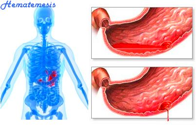
what is the best management for hematemsis of chronic liver failure patients ?


Hematemesis, or vomiting of blood, is a severe complication of chronic liver failure, and it requires immediate medical attention. The best management for hematemesis in chronic liver failure patients includes the following steps:


1- Stabilize the patient's vital signs and provide supportive care, such as oxygen therapy and intravenous fluids.


2- Administer medications to control bleeding, such as vasopressin or octreotide, which reduce the blood flow to the bleeding site.


3- Perform an upper gastrointestinal endoscopy to identify the source of bleeding and apply appropriate treatment, such as endoscopic band ligation, sclerotherapy, or balloon tamponade.


4- If endoscopic therapy fails to control bleeding, consider transjugular intrahepatic portosystemic shunt (TIPS) placement or surgical intervention, such as portacaval shunt or liver transplantation.


5- Prevent future bleeding episodes by addressing the underlying cause of chronic liver failure, such as alcohol abuse, viral hepatitis, or non-alcoholic steatohepatitis (NASH).


6- Ensure the patient receives ongoing medical care, such as regular monitoring of liver function tests, nutritional support, and management of complications, such as ascites, encephalopathy, or hepatorenal syndrome.


In summary, the management of hematemesis in chronic liver failure patients requires a multidisciplinary approach, involving gastroenterologists, hepatologists, critical care specialists, and transplant surgeons. The treatment plan should be tailored to the individual patient's needs and underlying cause of liver failure.

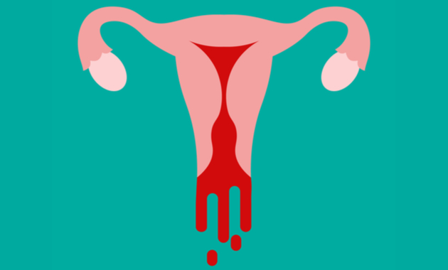
what is the best managment for vaginal bleeding after above knee amputation ?


Vaginal bleeding after above knee amputation is not a typical complication related to the surgery. It is crucial to determine the cause of the bleeding to provide the appropriate management. Some possible causes include trauma during surgery, coagulopathy (blood clotting disorder), or an unrelated gynecological issue.


If the bleeding is severe or persistent, the patient should seek immediate medical attention. The following steps may be taken to manage vaginal bleeding after an above-knee amputation:


1- Evaluate the patient's medical history and perform a physical examination to determine the cause of the bleeding.


2- Provide supportive care, such as ensuring the patient is well-hydrated and maintaining stable vital signs.


3- Administer medications, such as tranexamic acid or desmopressin, to control bleeding if necessary.


4- If the bleeding is due to a gynecological issue, refer the patient to a gynecologist for further evaluation and management.


5- If the bleeding is due to a coagulopathy, provide appropriate blood products and consult with a hematologist for further management.


Overall, the management of vaginal bleeding after above-knee amputation should be tailored to the individual patient's needs and underlying cause. It is essential to involve a multidisciplinary team of healthcare providers, including surgeons, gynecologists, and hematologists, to ensure optimal patient care.

Outlines of American trypanosomiasis ( chagas' disease )


Introduction:

Chagas disease, also known as American trypanosomiasis, is a parasitic infection caused by the protozoan Trypanosoma cruzi (T. cruzi). The disease is endemic in Central and South America, where it affects approximately 6 to 7 million people, with an additional 70 million individuals at risk of infection. The disease is transmitted to humans via the feces of triatomine bugs, also known as “kissing bugs”. The acute phase of the disease is usually mild or asymptomatic, but if left untreated, the chronic phase can lead to serious cardiac and digestive complications.


Etiology and Transmission:

T. cruzi is a hemoflagellate protozoan that is primarily transmitted to humans via the feces of infected triatomine bugs. These bugs typically live in the cracks and crevices of poorly constructed housing, and feed on the blood of sleeping humans at night. During feeding, the bug defecates near the site of the bite, and the feces, which contain the T. cruzi parasites, can then enter the human body through mucous membranes or broken skin. In addition to vector-borne transmission, Chagas disease can also be transmitted via blood transfusions, organ transplantation, or from mother to child during childbirth.


Clinical Manifestations:

The clinical manifestations of Chagas disease can be divided into acute and chronic phases. The acute phase usually lasts for several weeks and is characterized by mild or asymptomatic symptoms, including fever, headache, muscle pain, and swollen lymph nodes. In some cases, patients may develop a characteristic swelling of the eyelid on the side of the bite, known as a “RomaƱa sign”. However, in approximately 30% of cases, the acute phase can progress to a severe form of the disease, with myocarditis, meningoencephalitis, and other serious complications.


If left untreated, the chronic phase of Chagas disease can develop decades after initial infection, and can lead to severe cardiac and digestive complications. Cardiac complications are the most common and include cardiomyopathy, arrhythmias, and heart failure. Digestive complications can include megacolon and megaesophagus, which can lead to difficulty swallowing, malnutrition, and weight loss.


Diagnosis and Treatment:

Diagnosis of Chagas disease can be challenging, particularly in the chronic phase, as symptoms may be nonspecific and laboratory tests may have low sensitivity and specificity. Serological tests, such as ELISA and indirect immunofluorescence, are the primary diagnostic tests, although PCR and microscopy may also be used.


Treatment of Chagas disease is most effective in the acute phase, with antiparasitic drugs such as benznidazole and nifurtimox. These drugs are less effective in the chronic phase, and treatment is usually focused on managing symptoms and preventing complications. In cases of severe cardiac or digestive complications, surgery may be necessary.


Prevention:

Prevention of Chagas disease primarily involves control of the triatomine bug population, through measures such as improved housing, insecticide spraying, and bed net use. Blood transfusion and organ transplantation screening programs have also been implemented in endemic areas to reduce the risk of transmission through these routes.


Conclusion:

Chagas disease is a significant public health problem in Central and South America, with millions of individuals affected and at risk of infection. The disease is transmitted primarily by the triatomine bug, and can lead to serious cardiac and digestive complications if left untreated. Diagnosis and treatment can be challenging, particularly in the chronic phase, and prevention is focused on controlling the vector population and reducing transmission through blood transfusions and organ transplantation. Further research is needed to develop effective treatments for the chronic phase of the disease.


Get more about chagas' disease

Outlines of African Trypanosomiasis (African Sleeping Sickness)


Abstract:


African trypanosomiasis, also known as sleeping sickness, is a neglected tropical disease caused by the protozoan parasites Trypanosoma brucei. The disease is transmitted to humans and animals by the tsetse fly, a bloodsucking insect found only in sub-Saharan Africa. African trypanosomiasis is characterized by two distinct stages: an early hemolymphatic stage and a late meningoencephalitic stage, which can be fatal if left untreated. Diagnosis of African trypanosomiasis is challenging, and treatment options are limited, with many drugs having toxic side effects. There is an urgent need for new diagnostic tools and treatments for this disease.

Introduction:


African trypanosomiasis is a parasitic disease that affects humans and animals in sub-Saharan Africa. The disease is caused by the protozoan parasites Trypanosoma brucei, which are transmitted by the tsetse fly. There are two subspecies of T. brucei that cause human African trypanosomiasis: T. b. gambiense, which is responsible for over 95% of cases, and T. b. rhodesiense, which accounts for the remaining cases.

Epidemiology:


African trypanosomiasis is endemic in 36 countries in sub-Saharan Africa, where an estimated 69 million people are at risk of infection. The disease is most prevalent in rural areas where there is a high density of tsetse flies. The World Health Organization (WHO) estimates that there were approximately 9779 cases of African trypanosomiasis in 2020, with 92% of cases reported in the Democratic Republic of the Congo.

Clinical features:


The clinical features of African trypanosomiasis can be divided into two distinct stages: an early hemolymphatic stage and a late meningoencephalitic stage. In the early stage, patients may experience non-specific symptoms such as fever, headache, joint pain, and malaise. As the disease progresses to the late stage, patients may develop neurological symptoms such as confusion, behavioral changes, and sleep disturbances. The late stage is also characterized by the presence of trypanosomes in the cerebrospinal fluid, which can lead to coma and death if left untreated.

Diagnosis:


Diagnosis of African trypanosomiasis can be challenging, as the clinical symptoms are non-specific and can be similar to those of other diseases. The diagnosis is usually confirmed by the detection of trypanosomes in blood or cerebrospinal fluid using microscopy or other diagnostic tests such as the card agglutination test for trypanosomiasis (CATT).

Treatment:


Treatment options for African trypanosomiasis are limited, and many drugs have toxic side effects. The drugs used for the treatment of the early stage of the disease include pentamidine and suramin, while the drugs used for the late stage include melarsoprol and eflornithine. However, these drugs have limitations, and the emergence of drug-resistant trypanosomes is a major concern. There is an urgent need for new drugs with better efficacy and safety profiles.

Prevention and control:


Prevention and control of African trypanosomiasis rely on a combination of strategies, including the control of tsetse fly populations, the use of insecticide-treated bed nets, and the screening of blood donors. The WHO has set a goal to eliminate African trypanosomiasis as a public health problem by 2030, and progress is being made towards achieving this goal.


Get more about African Trypanosomiasis